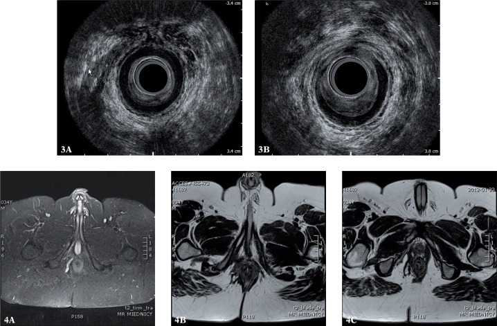
Figs. 3, 4.
Suprasphincteric fistula on the right wall in AES and MRI (3 A, 4 A) with the internal opening localized medially and anteriorly (3 B, 4 B). AES failed to identify a high track within the right branch of the puborectalis muscle (4 C). The defect of 1/3 of the anterior circumference of the internal sphincter in the medial part of the canal is better visible in AES than in MRI (3 B)

