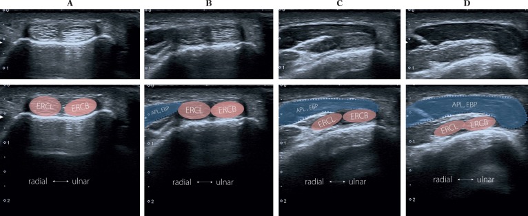
Fig. 7.
Second compartment. The probe is placed transversally as it is shown in Fig. 6. The tendons of the second compartment can be identified (tendon of extensor carpi radialis longus; ECRL, tendon of extensor carpi radialis brevis, ECRB). As the probe is moved proximally along the tendons of the second compartment (from position A to position D in Fig. 4), the muscles of the first compartment can be seen (abductor pillicis longus, APL; extensor pollicis brevis; EPB). Note how the muscles of the first compartment encroach superficially to the ECRL and ECRB (II)

