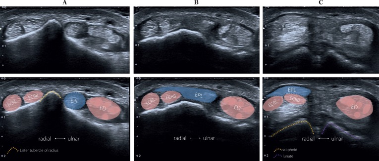
Fig. 9.
Third compartment (dorsal wrist) with the extensor pollicis longus tendon (EPL). The probe is placed transversally as it is shown in Fig. 8, point A. It is subsequently moved distally up to the EPL insertion. A – the “starting position”: the EPL tendon is situated on the ulnar side of the Lister tubercle. B and C show the EPL tendon encroaching superficially from the tendons of the second compartment (tendon of the extensor carpi radialis longus; ECRL, tendon of the extensor carpi radialis brevis, ECRB) to the tendon of the extensor digitorum communis which belongs to the fourth compartment

