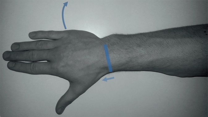
Fig. 15.
Scapholunate ligament (dorsal portion) – scanning technique. The probe should be placed transversally on the level of the Lister tubercle and then swept distally. The curved arrow demonstrates the ulnar deviation of the wrist – it is useful in the evaluation of the echostructure and integrity of the dorsal part of the scapholunate ligament

