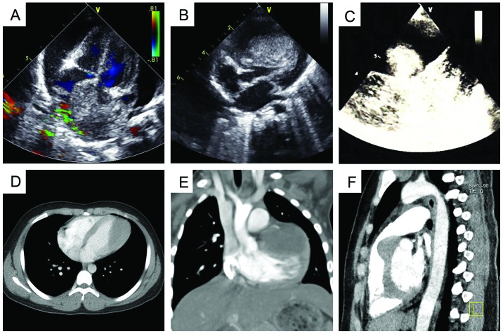
Figure 1.
Echocardiography and computed tomography angiography prior to surgery. (A) Tumor in the atrial septum with moderate pericardial effusion. (B) Right ventricular tumor with outflow tract obstruction. (C) Intrapericardial mass. (D) Left ventricle tumor. (E) Left ventricle tumor with left coronary artery involvement. (F) Right ventricular tumor.

