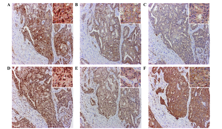
Figure 1.
Representative immunostaining for MT-2A, E-cadherin, IL-6, cyclin E, PCNA and Bcl-2 in consecutive sections of a PCa specimen. Expression and location of different proteins in PCa cells: (A) MT-2A in cytoplasm and nuclei; (B) E-cadherin in the cytoplasm; (C) IL-6 in the cytoplasm; (D) cyclin E in the nuclei; (E) PCNA in the nuclei; and (F) Bcl-2 in the cytoplasm. Magnification, ×100; magnification of insets, ×200. MT-2A, metallothionein-2A; IL-6, interleukin 6; PCNA, proliferating cell nuclear antigen; Bcl-2, B cell lymphoma 2; PCa, prostate cancer.

