
Fig. 15.
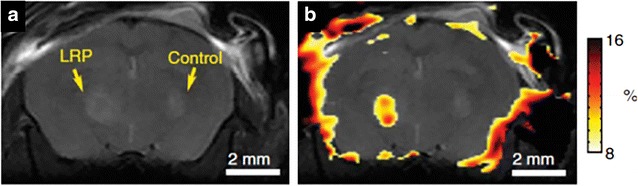
Lysine rich protein (LRP) based MR reporter genes transfected with the 9L gliosarcoma cells before implantation in the rat brain. On anatomical imaging both the LRP and control xenografts show the similar signal intensity (a). The CEST map highlighted the LRP xenograft due to the expression of lysine rich protein, which can be easily detected through APT CEST (b). Reproduced with permission from the Nature Publishing Group and Gilad et al. [112]