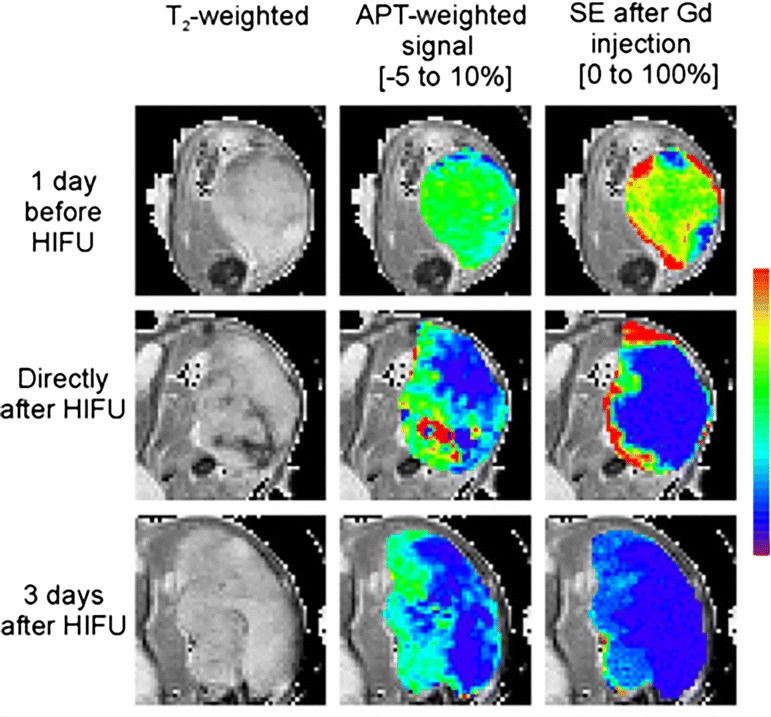
Fig. 7.

Monitoring HIFU treatment in cancer. Proton anatomical image, APT weighted image and GD contrast enhanced image show the changes in tumor following HIFU treatment. Decreased APT contrast following HIFU was observed which is comparable to the Gd based contrast enhancement study. Reproduced with permission from John Wiley and Sons and Hectors et al. [91]
