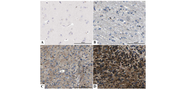
Figure 1.
Immunohistochemical staining of IF1 in glioma and NB tissues. IF1 was localized within the cytoplasm. (A) NB tissues without IF1 expression. (B) Low, (C) medium and (D) high expression of IF1 in the tumor cells of glioma tissue compared with (A). Scale bar, 100 µm. NB, normal brain; IF1, adenosine triphosphatase inhibitory factor 1.

