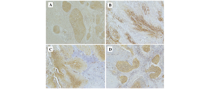
Figure 1.
Representative images of the immunohistochemical visualization of (A) hypoxia inducible factor-1α, (B) carbonic anhydrase-IX, (C) glucose transporter-1 and (D) vascular endothelial growth factor in cervical squamous cell carcinoma. Fixed cells were stained with specific antibodies and horseradish-peroxidase secondary antibodies, and then counterstained with hematoxylin (magnification, ×200).

