Figure 1.

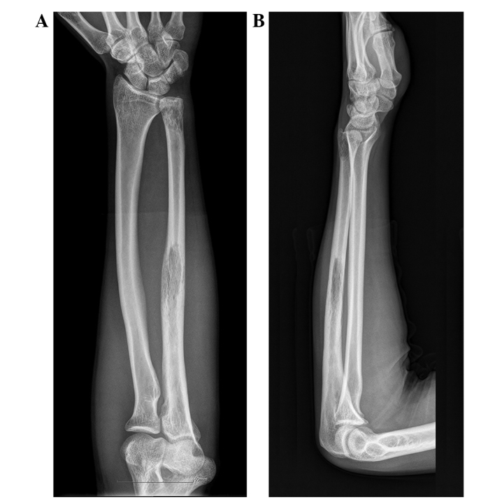
(A) Anteroposterior and (B) lateral plain radiographs showing an osteolytic lesion with cortical destruction in the proximal, middle and distal ulna.

(A) Anteroposterior and (B) lateral plain radiographs showing an osteolytic lesion with cortical destruction in the proximal, middle and distal ulna.