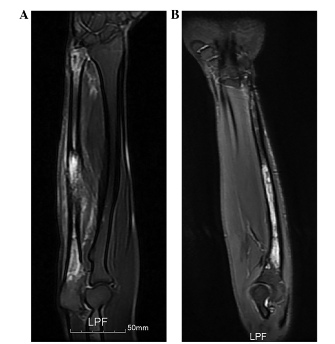
Figure 2.

Magnetic resonance imaging findings (T2-weighted). (A) Prior to neoadjuvant chemotherapy. The intramedullary tumor involved nearly the full length of the ulna, with the exception of the proximal olecranon. The surrounding cortex was partially involved, and the soft-tissue components around the tumor appeared patchy and hyperintense. (B) Following neoadjuvant chemotherapy. The tumor regressed following two courses of intensive chemotherapy.
