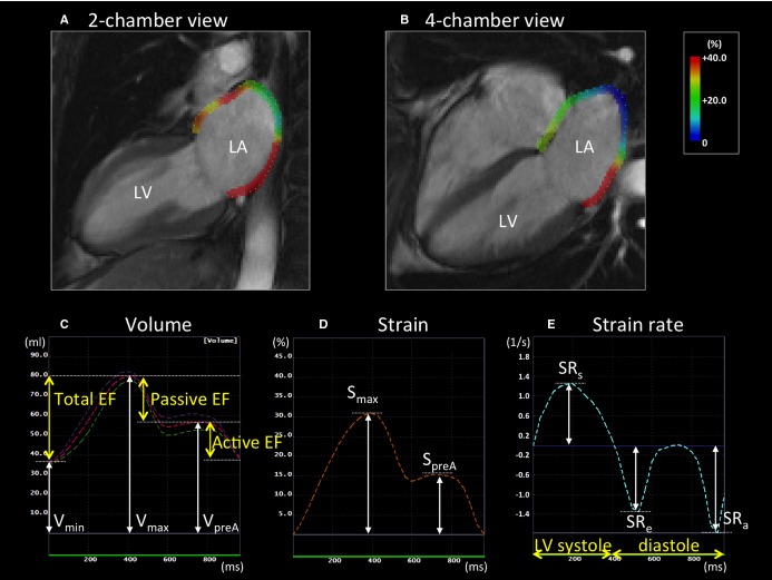
Figure 2.
LA measurements by tissue‐tracking CMR in a patient without stroke. A and B, Left atrium (LA) longitudinal strain in the 2‐ and 4‐chamber views at the end of left ventricular (LV) systole. C, LA volume curve. The pink dotted line is the average of the values of volume in the 2‐ and 4‐chamber views. The LA maximum volume (Vmax), the pre‐atrial contraction volume (VpreA), and the minimum volume (Vmin) were identified. The LA emptying fractions (EFs) were calculated using Vmax, VpreA, and Vmin. D and E, The LA strain and strain rate curve. The LA maximum strain (Smax) and pre‐atrial contraction strain (SpreA) were identified from the strain curve. The strain rates during LV systole (SRs), LV early diastole (SRe), and atrial contraction (SRa) were also analyzed from the strain rate curve. CMR indicates cardiac magnetic resonance.

