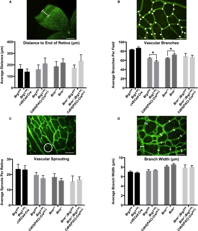
Figure 2.
SWI/SNF mutants do not display major vascular defects in the neonatal retina. Retinas from postnatal day 7 (P7) pups were stained with isolectin B4 and flat mounted to visualize the vasculature. A, Distance (μm) was measured from the edge of the vascular front to the end of the retina (white bar). Four measurements were made for each retina. B, Retina vascular branches (white dots) were counted in 10 fields for each retina. C, Vascular sprouts (white circle) were counted in 10 fields for each retina. D, Branch widths were measured from 10 vessels in 10 different fields for each retina. For A through D, pictures show representative measurement criteria. Data represent averages±SEM from 8 to 11 animals for each genotype. *P<0.05; Student t test. Scale bars=500 μm (A); 50 μm (B through D). Brg1 indicates Brahma‐related gene 1; Brm, Brahma; SWI/SNF, SWItch/Sucrose NonFermentable.

