Figure 3.

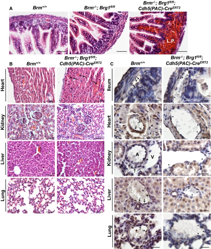
Characterization of Brm−/−;Brg1fl/fl;Cdh5(PAC)‐CreERT2 infant mice. A, P14 Brm+/+ (control), Brm−/−;Brg1fl/fl (control), and Brm−/−;Brg1fl/fl;Cdh5(PAC)‐CreERT2 (double‐mutant) ilea were hematoxylin and eosin stained. Hemorrhage is seen in the lamina propria (L.P.) of the double‐mutant ileum. B, Control and double‐mutant tissues from postnatal day 14 (P14) pups were stained with hematoxylin and eosin. Top images show hemorrhage in the intramuscular tissue of the double‐mutant heart (arrows). No hemorrhage was detected in double‐mutant kidney, liver, or lung tissues. C, Control and double‐mutant tissues from P14 pups were immunostained for BRG1 (brown) and counterstained with hematoxylin (blue). Arrows point to endothelial cells that express BRG1. Asterisks highlight endothelial cells where BRG1 expression is diminished. Scale bars=50 μm (A and B); 100 μm (C). A indicates artery; Brg1, Brahma‐related gene 1; Brm, Brahma; V, vein.
