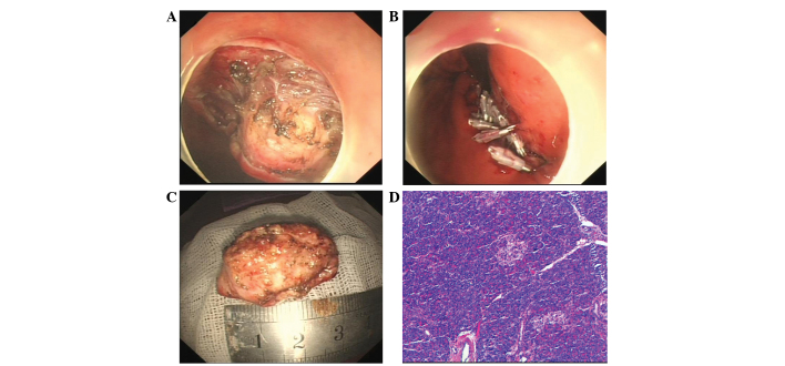
Figure 2.
Endoscopic submucosal dissection (ESD) of the ectopic pancreas. (A) Endoscopic direct image showing a soft, yellow tumor. (B) Nylon loop with clips used to suture the mucosal defects and perforation of the stomach on endoscopy. (C) The inner surface of the resected specimen. (D) Histopathological findings revealing only acinar cells in the tissue sample obtained by the bloc biopsy (hematoxylin and esoin staining; magnification, x100).

