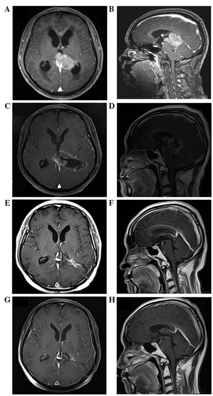
Figure 1.

MRI in diagnosis and follow-up assessment of pineoblastoma. Images of the ventricle and pineal gland were captured in the axial (left column) and sagittal (right column) planes. (A and B) Initial preoperative MRI showed a massive tumor measuring 4.1×5.1×3.9 cm in the pineal region. It was heterogeneously enhanced following gadolinium administration. The tumor was close to the midbrain and thalamus, and protruded forward into the posterior part of the third ventricle and backward into the cistern of the great cerebral vein. Both internal cerebral veins and the great cerebral vein were enclosed within the tumor. It compressed the triangular region of the left lateral ventricle from its lower and medial side and mesencephalic aqueduct from its posterior side, and caused obstructive hydrocephalus in the supratentorial ventricles. (C and D) Immediate postoperative MRI. A residual mass with gadolinium enhancement in pineal region was observed. (E and F) MRI at 2 months after radiotherapy; complete regression of the tumor was observed following radiotherapy. (G and H) MRI at 36 months after irradiation; no evidence of tumor recurrence was observed. MRI, magnetic resonance imaging.
