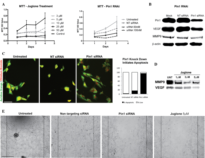
Figure 1.
(A) Graphs revealing the reduction in proliferation caused by Pin1 siRNA and juglone in glioblastoma cells in vitro. The difference between juglone-treated and -untreated groups and siRNA-treated versus non-targeting siRNA or untreated groups was significant (P<0.05). (B) Immunoblotting revealing the reduction in the levels of Pin1, VEGF and MMP9 by Pin1 siRNA. (C) Pin1 siRNA transfection activated apoptosis in the glioblastoma cells, whereas the non-targeting siRNA-treated and control cells were healthy. Acridine orange/ethidium bromide vital staining reveals apoptotic cells with red and orange dots in the perinuclear region, healthy cells in green and necrotic cells in red. In total, 95% of the cells that were counted in the Pin1 siRNA group were apoptotic (n=115) (scale bar, 200 µm). (D) A significant reduction in VEGF and MMP9 levels observed following juglone treatment is shown. (E) Wound healing assay revealing that Pin1 siRNA and juglone were effective at disrupting the migration ability of glioblastoma cells (scale bar, 50 µm). Pin1, peptidyl-prolyl cis/trans isomerase NIMA-interacting 1; siRNA, small interfering RNA; VEGF, vascular endothelial growth factor; MMP9, matrix metalloproteinase 9; RNAi, RNA interference; NT siRNA, non-targeting siRNA; UNT, untreated; OD, optical density.

