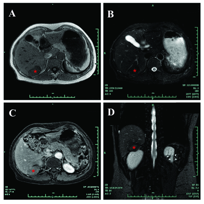
Figure 2.
MRI confirmed the presence of a solid neoplasm (asterisk), with (A) low signal intensity on T1-weighted images and (B) high signal intensity on T2-weighted images. (C) The signal intensity of the neoplasm was slightly enhanced on the contrast-enhanced phases of dynamic MRI. (D) Coronal T2-weighted images.

