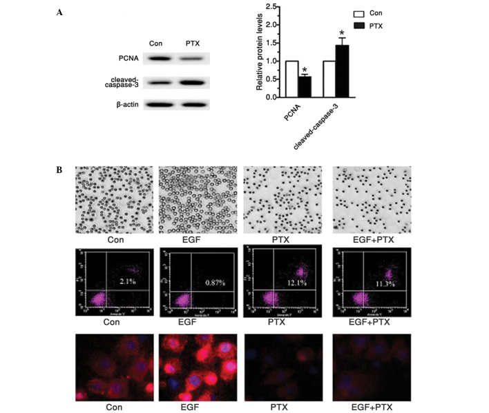
Figure 4.
PTX suppressed tea8113 cell proliferation and induced its apoptosis. (A) Western blot analysis revealed decreased proliferation marker, proliferative cell nuclear antigen, and enhanced cleaved-caspase-3 levels. (B) Microscope, flow cytometry and immunofluorescence were applied to determine cell numbers, apoptosis and epidermal growth factor receptor protein levels. *P<0.05 versus control.

