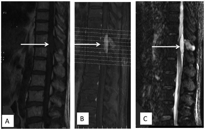
Figure 1.
(A) Sagittal T1-weighted image prior to gadolinium infusion showing hypointense signals in the spinous process of the T11 vertebra and an epidural mass with hypointense signals at the T11 and T12 vertebral level. (B) Sagittal T2-weighted, fat suppressive magnetic resonance imaging revealing hyperintense signals in the spinous process of the T11 vertebra and an epidural mass with hyperintense signals at the T11 and T12 vertebral level. (C) Well-enhanced mass following gadolinium-DTPA infusion and compression of the thoracic spinal cord.

