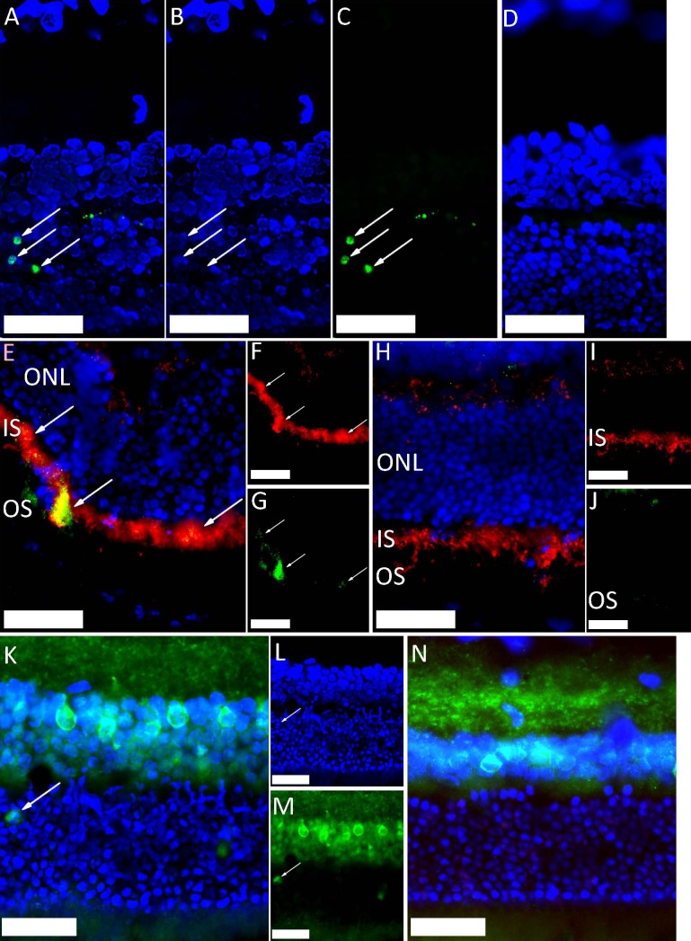
Figure 1.
Immunohistochemical staining of rat outer retinal layers, peripheral to the impact site, after ballistic injury (DAPI-stained nuclei are shown in blue [scale bar: 50 μm]). (A–D) TUNEL-stained photoreceptor nuclei, in green (arrows) at 2 days after injury ([A] blue and green channels [B, C], respectively). TUNEL-negative uninjured control retina (D). (E–J) Caspase-9-stained green (arrows) in photoreceptor inner segments (E, G). Inner segments stained red with anticomplex IV antibody (E, F) at 5 hours after injury. Uninjured control retina (H–J) did not caspase-9 staining. (K–N) Increased caspase-9 levels in photoreceptor cell bodies (arrow) are shown at 2 days in injured retina (K–M). Uninjured control retina did not show caspase-9 staining in the ONL (N).

