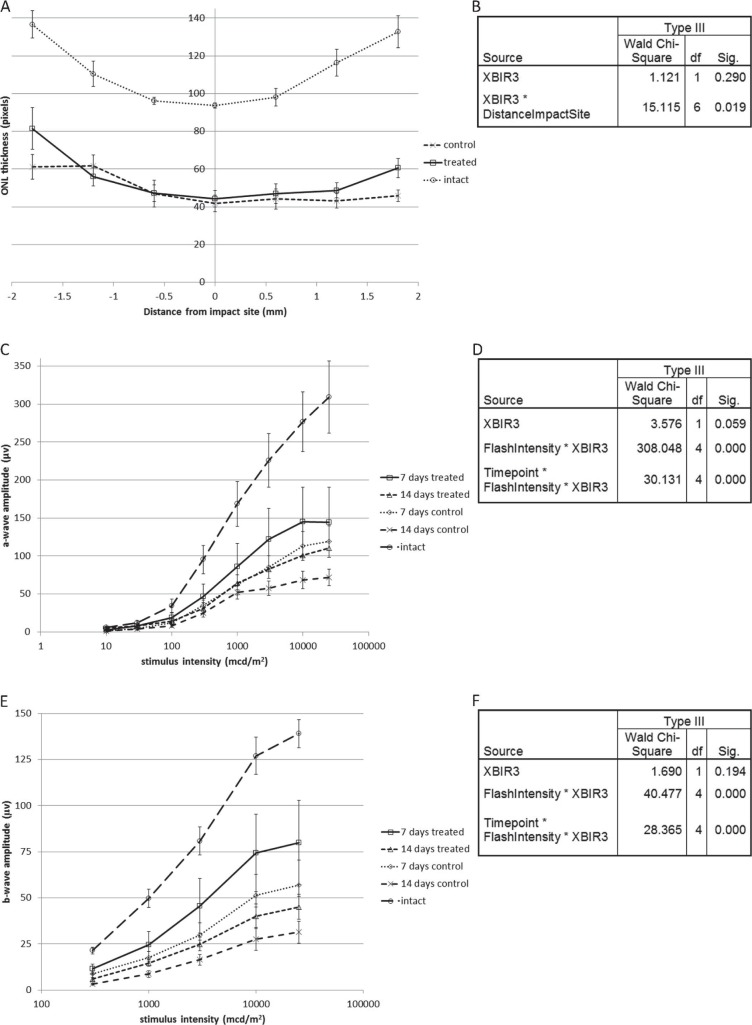
Figure 3.
(A) ONL thickness with and without intravitreal Pen-1–XBIR3 is shown as a function of distance from the impact site. ONL thickness in intact controls is shown for comparison. (B) Test of model effects after analysis of ONL thickness, including treatment with Pen-1–XBIR3, distance from the impact site and a term to model their interaction, displaying relevant terms only. (C) Scotopic a-wave amplitude after injury as a function of stimulus intensity with and without intravitreal Pen-1–XBIR3 is shown. Scotopic a-wave amplitude in intact controls is shown for comparison. (D) Test of model effects after analysis of scotopic a-wave amplitude including time after injury (Timepoint), stimulus intensity (FlashIntensity), Pen-1–XBIR3 compared to Pen-1 alone (XBIR3), and terms to model all 2- and 3-way interactions, displaying relevant terms only. (E) Photopic b-wave amplitude after injury as a function of stimulus intensity with and without intravitreal Pen-1–XBIR3 is shown. Photopic b-wave amplitude in intact controls is shown for comparison. (F) Test of model effects is shown after analysis of photopic b-wave amplitude, including time after injury, stimulus intensity, treatment, and terms to model all 2- and 3-way interactions, displaying relevant terms only. Caspase-9 inhibition by Pen-1–XBIR3 increased scotopic a- and photopic b-wave amplitudes with an effect that was more pronounced at higher stimulus intensities and at 14 than at 7 days after injury. Caspase-9 inhibition also increased ONL thickness peripherally but not centrally to the impact site. All data are means ± standard errors of the mean.

