
Figure 3.
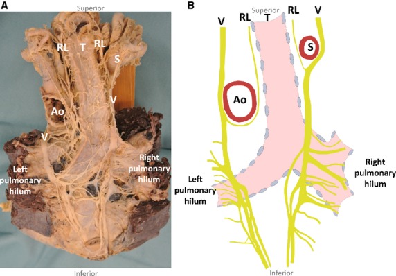
Schematic drawing of the right and left posterior pulmonary plexuses from a posterior view (B) with the corresponding photograph (A). Ao, aorta; RL, recurrent laryngeal nerve; S, subclavian artery; T, trachea; V, vagus nerve.
Official websites use .gov
A
.gov website belongs to an official
government organization in the United States.
Secure .gov websites use HTTPS
A lock (
) or https:// means you've safely
connected to the .gov website. Share sensitive
information only on official, secure websites.
Schematic drawing of the right and left posterior pulmonary plexuses from a posterior view (B) with the corresponding photograph (A). Ao, aorta; RL, recurrent laryngeal nerve; S, subclavian artery; T, trachea; V, vagus nerve.