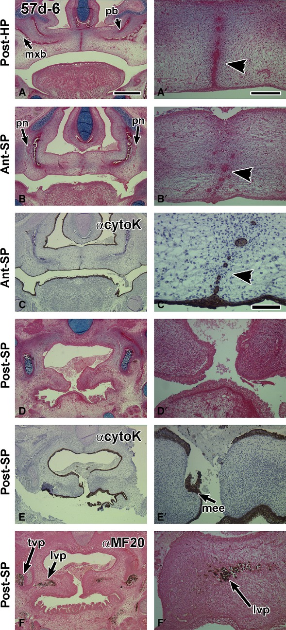
Figure 2.

Frontal sections through a 57-day fetus with a midline epithelial seam in the hard palate while in the soft palate the seam is degrading. (A,A′) A robust medial edge seam in the hard palate that is intact orally (arrowhead) and breaking down nasally. (B,B′) The border with the soft palate is identified by the presence of the palatine nerve lateral to the palatine bones. The midline appears to have epithelial islands (arrowhead). (C,C′) The pan-cytokeratin antibody identifies clearly that there are epithelial remnants in the midline (arrowhead in C′). The antibody also stains the dorsum of the tongue and pharynx. (D,D′) A posterior section through the open soft palate. (E,E′) an adjacent section to (D) stained with anti-cytokeratin. The medial edge epithelium is torn, which is an artifact of specimen preparation. (F,F′) Myosin heavy chain antibody MF-20 stains the palate musculature. lvp, levator veli palatini; mee, medial edge epithelium; mxb, maxillary bone; pb, palatine bone; pn, palatine nerve; t, tongue; tvp, tensor veli palatini. Scale bars: (A) 1 mm, (A′) 200 μm and applies to B′,D′,E′,F′; (C′) 100 μm.
