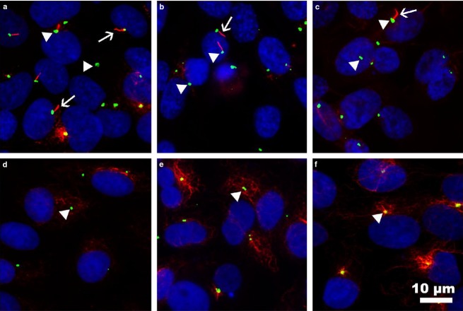
Figure 6.
Immunofluorescence staining of primary cilia in human and rat follicular cell lines. Primary cilia were studied in human Nthy-ori 3-1 (a,f), FTC-133 (b), and 8505C (c) and rat PC-Cl3 (d), and FRT (e) cell lines by staining the axonemes with anti-acetylated α-tubulin (red) and the basal bodies with anti-γ-tubulin (green). Nuclei were labeled with DAPI. Primary cilia were present in approximately 43% of normal human follicular cells (Nthy-ori 3-1), in 21% of neoplastic human FTC-133 cells and in 7% of anaplastic 8505C cells, but were absent in all the rat cell lines tested (PC-Cl3, FRT) and non-starved human control Nthy-ori 3-1 cells (f).

