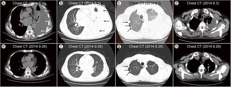
Fig. 1.

Chest CT (2014.6.3) showed a left-sided pleural effusion with compressive atelectasis of the left lower lobe (a, b), multiple nodules scattered in both lungs (c), and calcification in the nodules in the left upper lobe (d). Chest CT (2014.9.26) revealed that the left pleural effusion was completely resolved (e, f), but multiple nodules and calcification were still present (g, h)
