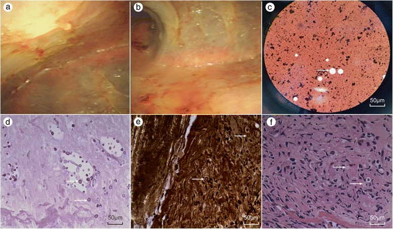
Fig. 2.

Intuitive thoracoscopy revealed localized pleural adhesion and a diffuse, cellulose-like pleura (a, b). Cryptococcus neoformans was identified by direct India ink staining of the cerebrospinal fluid (CSF) (c). Periodic acid-Schiff stain (PAS) and methenamine silver stain revealed numerous Cryptococcus organisms in pleural sections (d, e). Re-examination of previous sections revealed Cryptococcus-like organisms that had been previously overlooked, and the capsule was not stained (f)
