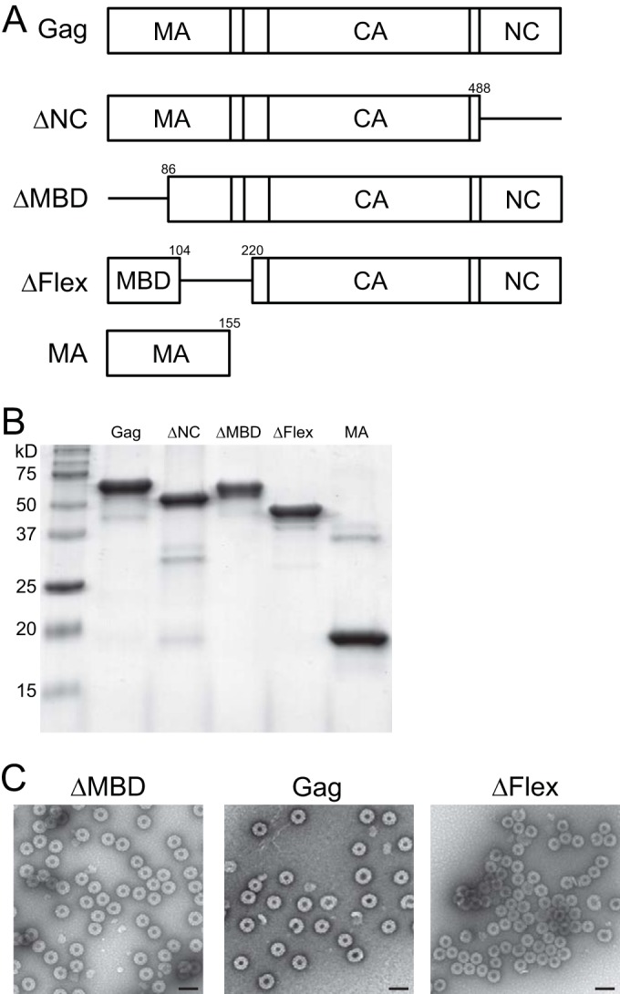
FIG 1.

Proteins used in this study. (A) Schematic representation of proteins. Note that the proteins MBD and GST-NC are not shown. Numbers indicate amino acid positions. (B) Stained gel of the proteins used in liposome binding assays. For analysis by size exclusion chromatography (SEC), small angle X-ray scattering (SAXS), and neutron reflectometry (NR), the proteins were further purified by SEC. (C) Virus-like particles (VLPs) assembled in vitro and imaged by negative-stain electron microscopy. Scale bar, 100 nm.
