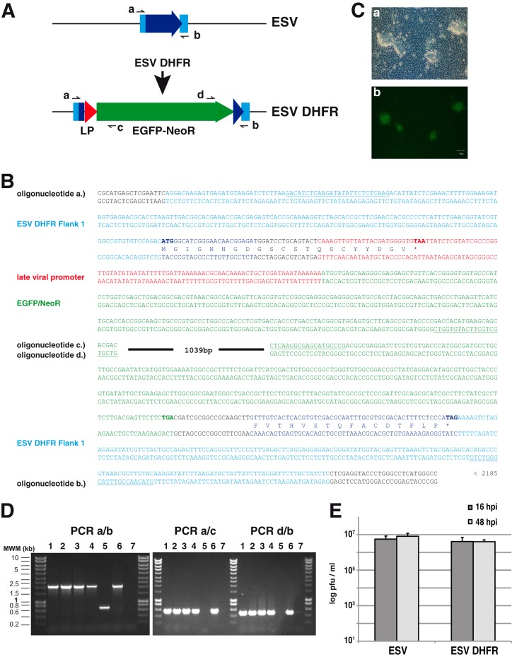
FIG 3.
Method for the generation of recombinant ESV using homologous recombination. (A) Schematic representation of the genomic structure of the parental ESV and the ESVΔDHFR deletion mutant generated indicating the fragmented locus, the late viral promoter, and the neomycin resistance/EGFP fusion cassette used for selection, as well as the positions of the oligonucleotides used for PCR analyses. (B) Relevant sequences of a fragment of the plasmid used for homologous recombination, representing the genomic sequence of the recombinant virus in this area. The sequence of the viral promoter corresponding to the 92 bp upstream of the initiation codon of Singapore grouper iridovirus ORF 125R is shown in red. The complete sequence of the plasmid is available upon request. The amino acids corresponding to the putative ESV DHFR are shown in blue, and translation frames are indicated. (C) Bright-field (a) and fluorescence microscopy (b) of EPC cells showing developing ESVΔDHFR replication plaques at 24 hpi. (D) PCR analyses using the indicated oligonucleotide pairs of purified DNA derived from four individual recombinant ESV clones (lanes 1 to 4) and the parental ESV (lane 5), along with a positive (plasmid used for the generation of the recombinant virus (lane 6) and negative (no-DNA) control (lane 7). MWM, molecular weight markers. (E) ESV and ESVΔDHFR virus yields at the indicated time points after high-multiplicity infection of EPC cells.

