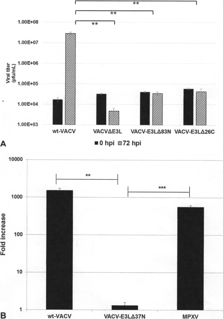
FIG 5.
Replication in JC cells. (A) JC cells were infected with the indicated viruses at an MOI of 0.01 PFU/cell. Infected cells were harvested at 0 and 72 hpi, and the titer was determined by plaque assay in RK-E3L cells. Data are presented as means with standard errors from three experiments. **, P ≤ 0.01. (B) JC cells were either mock infected or infected with wt VACV, VACV E3LΔ37N, or MPXV at an MOI of 0.01 PFU/cell. Viruses were harvested at 72 hpi, and the titer was determined by plaque assay in BSC-40 cells. Data represent the fold increase in viral growth at 72 hpi and are presented as means with standard errors from multiple experiments. Statistical analyses were done by using an unpaired t test comparing wt VACV and MPXV to VACV-E3LΔ37N. **, P ≤ 0.01; ***, P ≤ 0.001.

