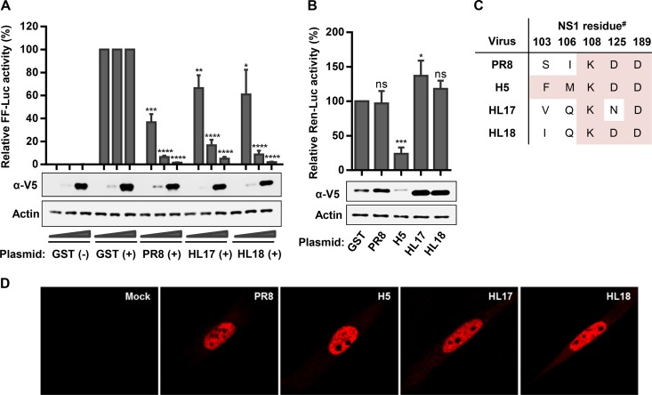
FIG 1.
Bat IAV NS1 proteins antagonize IFN-β induction but do not block general host gene expression. (A) Impact of bat IAV NS1s on IFN-β induction. 293T cells were cotransfected with increasing amounts of pLVX-based plasmids expressing a V5-tagged version of the indicated NS1 protein (or GST), together with an IFN-β promoter-driven firefly luciferase (FF-Luc)-expressing plasmid (p125luc [25 ng]) and an HSV-TK promoter-driven Renilla luciferase (Ren-Luc) plasmid (pRL-TK [25 ng]). Twenty-four hours posttransfection, cells were infected with SeV (+) or mock infected (−) for 16 h to stimulate the IFN-β promoter. Relative FF-Luc activity was determined as the ratio between FF- and Ren-Luc. Values were normalized to GST plus SeV (set to 100% for each GST dose). (B) Impact of bat IAV NS1s on general host gene expression. 293T cells were cotransfected with 0.5 μg V5-tagged GST or the indicated NS1 expression plasmid and 50 ng Renilla luciferase-expressing plasmid (pRL-TK). Twenty-four hours later, total Ren-Luc levels were determined, and values were normalized to GST (set to 100%). For panels A and B, bars represent means and standard deviations from three independent experiments. Lysates from parallel samples were analyzed by Western blotting for the indicated proteins and are shown from a representative experiment. Significance was calculated using the Student's t test comparing the appropriate GST dose with each respective NS1 dose (****, P < 0.0001; ***, P < 0.001; **, P < 0.01; *, P < 0.05; ns, not significant). (C) Lack of CPSF30-binding consensus residues in the bat IAV NS1 proteins. The table highlights the amino acid residues for each NS1 studied. #, residue numbering based on PR8 NS1 due to bat IAV NS1 amino acid insertions. Pink-shaded residues indicate CPSF30-binding consensus. (D) Intracellular localization of bat IAV NS1s in human lung fibroblasts. MRC5-hTERT cells were transfected with 0.5 μg of plasmid expressing the indicated V5-tagged NS1 protein or empty vector. Cells were fixed and permeabilized 24 h posttransfection, immunostained for the V5 tag, and visualized by confocal microscopy.

