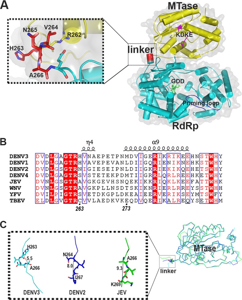
FIG 1.
(A) Crystal structure of DENV3 NS5 is displayed as a cartoon (PDB code 4V0R). The MTase domain is colored in yellow, the RdRp domain is in cyan, and linker residues 263 to 266 are in red. The inset shows a closeup view of the linker region. The KDKE catalytic tetrad of MTase is shown as magenta sticks and labeled; the GDD active site is in green, and the priming loop is in blue and labeled. (B) Sequence alignment of the linker regions between the MTase and RdRp domains of NS5 from DENV1 to -4 and various flaviviruses showing the secondary structures observed in DENV3 NS5. WNV, West Nile virus; YFV, yellow fever virus; TBEV, tick-borne encephalitis virus. (C) Superposition of the MTase domain of the flaviviral NS5 structures with structural information for the linker region. The distance between Cα's of the first and fourth linker residues was measured. DENV3 NS5, PDB code 4V0R; DENV2 MTase, PDB code 1L9K; JEV NS5, PDB code 4K6M.

