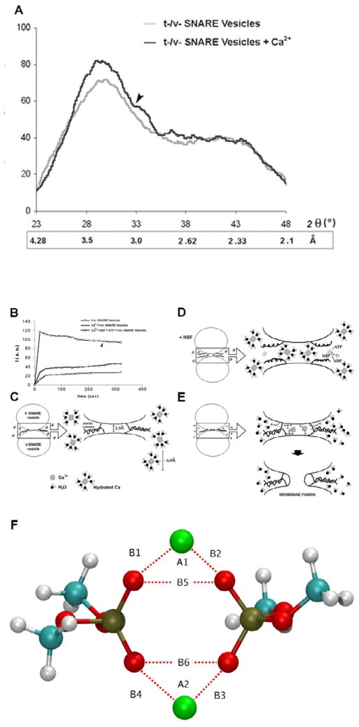
Figure 6.
(A) Wide-angle X-ray diffraction patterns of interacting t-SNARE and v-SNARE vesicles. Representative diffraction profiles from one of four separate experiments using t- and v-SNARE-reconstituted lipid vesicles, both in the presence or absence of 5 mM Ca2+ is shown. The arrow marks the appearance of a new peaks in the X-ray diffractogram, following addition of calcium. (B–E) Light scattering profiles of SNARE-associated vesicle interactions. (B,C) Addition of t-SNARE- and v-SNARE-vesicles in calcium-free buffer, lead to a significant increase in light scattering. Subsequent addition of 5 mM Ca2+ (marked by the arrowhead) has no significant effect on light scattering. (B,D) In the presence of NSF-ATP (1 μg/ml) in the assay buffer containing 5 mM Ca2+, significantly inhibits vesicle aggregation and fusion. Pi denotes inorganic phosphate. (B,E) When the assay buffer was supplemented with 5 mM Ca2+, prior to the addition of t- and v-SNARE vesicles, it led to a 4-fold decrease in light scattering intensity due to Ca2+-induced aggregation and fusion of t-/v-SNARE-apposed vesicles. Light scattering profiles shown are representatives of 4 separate experiments. (F) Ca2+-DMP− ring complex observed during molecular dynamics simulations. Atoms are colored as follows: Carbon (cyan), hydrogen (white), oxygen (red), phosphorous (gold), and Ca2+ (green). B1=2.146 Å, B2 = 2.145 Å, B3 = 2.140 Å, B4=2.145 Å, B5 = 2.92 Å, B6 = 2.92 Å, A1 = 85.79°, A2 = 85.07°.

