
Figure 1.
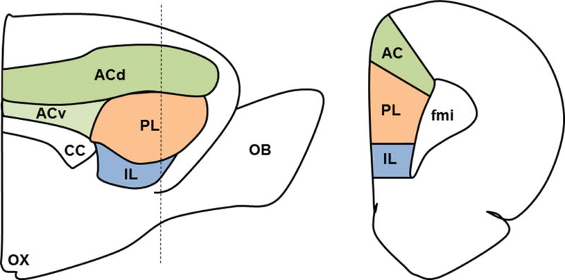
mPFC Neuroanatomy. Sagittal (left) and coronal (right) sections of the medial prefrontal cortex (mPFC) showing the anterior cingulate (AC), prelimbic (PL), and infralimbic (IL) cortices. The dotted line on the sagittal view indicates the location of the coronal section. Other abbreviations as follows: olfactory bulb (OB), forceps minor of the corpus callosum (FMI), corpus callosum (CC), dorsal AC (dAC), ventral AC (vAC), optic chiasm (OX).