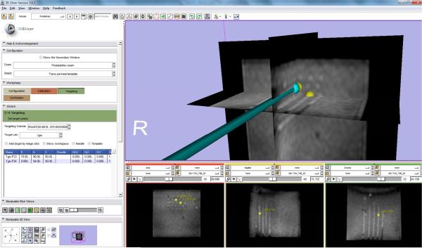
Fig. 9.
The clinical GUI integrated inside 3D Slicer shows a virtual needle whose position was streamed from control software, was inserted to move to the selected targets (shown as yellow spheres) during needle placement accuracy evaluation. The needle tracks were visualized inside the MRI volume. The first column of the bottom sub-figures shows the 5 × 5 needle tracks.

