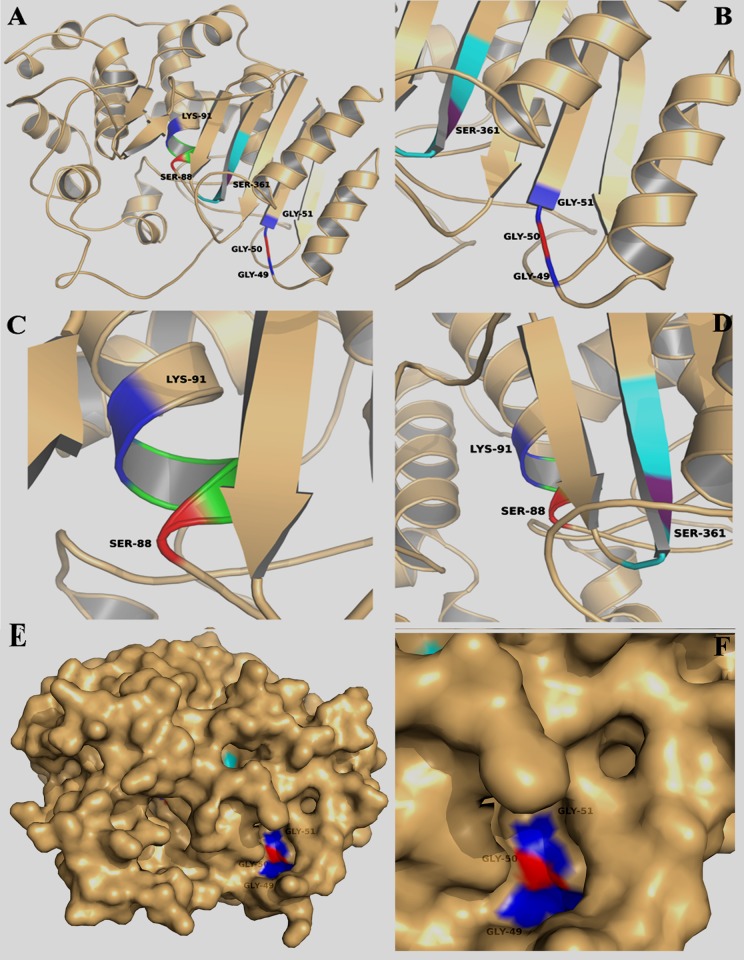
Fig 4. Homology model of LipL (template: EstA carboxylesterase, PDB code: 3ZYT).
(A) Ribbon view of LipL model which consisted of several α helices and β sheets. The predicted active residues are shown in different colors. (B) Enlarged view of GGG motif (residues G49 and G51 are shown in blue; residue G50 is shown in red). (C) The close-up view of the S-x-x-K motif (residue S88 is shown in red; residue K91 is shown in blue). (D) Enlarged view of the G-x-S-x-G motif (residue S361 is shown in purple). (E) The partially transparent surface representation of LipL. The GGG motif is highlighted shown in blue and red colors. It is noteworthy that the GGG motif is formed a catalytic pocket surface structure. (F) The close-up view of the surface representation of the GGG motif.

