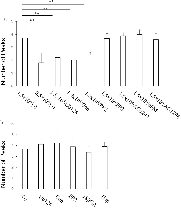
Fig 6. Pharmacological characterization of the calcium increase patterns of HeLa cells in response to histamine.
(a) Effects of long-term pharmacological treatments during cell culture. Cells were seeded at 1.5 x 10⁴ cells/cm2 or 0.5 x 10⁴ cells/cm2, and cultured in the presence or absence or pharmacological agents, including 1 μM U0126, 30 μM Genistein (Gen), 10 μM PP2, 10 μM PP3, 10 μM AG1476, 10 μM AG1247 and 5 μg/ml anti-bFGF neutralizing antibody (bFM) for 48 h prior to calcium imaging experiments. Numbers of peaks of calcium increases during 2 min stimulation with 30 μM histamine were plotted. ** p < 0.01, n = 20 cells. (b) Effects of acute pharmacological treatments during calcium imaging. Cells were seeded at 1.5 x 10⁴ cells/cm2, cultured in normal medium for 48 h and subjected to calcium imaging. Each pharmacological treatment, including, U0126, Genistein, 10 μM 18 β-glycyrrhetinic acid (18βGA) and 2 mM heptanol (Hep) started 1 min prior to histamine stimulation. Numbers of peaks of calcium increases during 2 min stimulation were plotted. ** p < 0.01, n = 20 cells

