
Fig. 2.
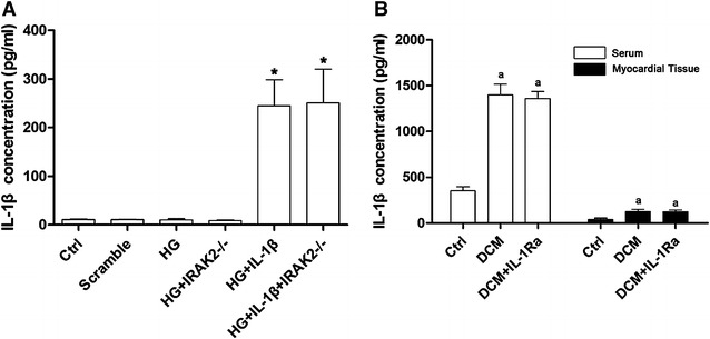
A Columns in this figure demonstrated the IL-1β level in supernatant of cultured primary myocytes by ELISA. According to different treatments, equal amount myocytes were divided into Ctrl (control), Scramble (cells transfected with scramble siRNA), HG (cells incubated by medium with glucose concentration at 33 mmol/L), HG + IRAK2−/− (cells treated with high-glucose medium and transfected with siRNA targeting irak2), HG + IL-1β (cells treated with high-glucose medium and exogenous IL-1β) and HG + IL-1β + IRAK2−/− (cells treated with high-glucose medium and exogenous IL-1β and transfected with siRNA against irak2) respectively [asterisk differences were significant when compared with HG (p < 0.05]. B Columns in this part showed the detected IL-1β in serum and myocardial tissue supernatant collected from normal control (Ctrl), DCM rats (DCM) and DCM rats received IL-1Ra administration (DCM + IL-1Ra) respectively [letter a differences were significant when compared with Ctrl (p < 0.05)]