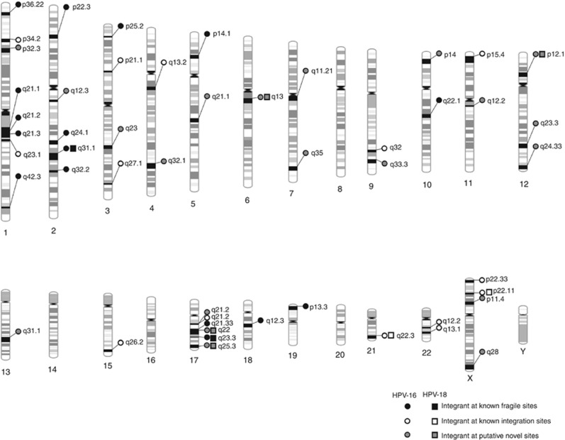
Figure 6.
Schematic representation of all HPV16 and 18 integration sites in the human genome detected across cervical cancer samples using HPVDetector. Site of integration as determined by HPVDetector in cervical cancer samples is shown. HPV16 integration sites are depicted by circles and HPV18 by rectangles. Black, open and grey circles or rectangles represent integrations at known fragile sites, at known integration sites and at novel sites, respectively.

