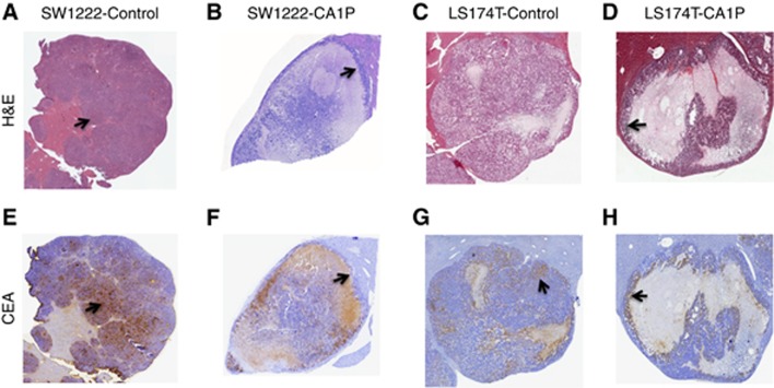
Figure 2.
Representative haematoxylin and eosin (A–D) and anti-CEA immunohistochemical (E–H) micrographs demonstrating the effect of CA1P on the distribution of viable cells and CEA in SW1222 (A, B, E, F) and LS174T (C, D, G, H) intrahepatic metastases. Control SW1222 metastases are almost totally viable (arrow, A), with relatively uniform expression of CEA (arrow, E). After treatment with CA1P, viable CEA-expressing tumour cells are restricted to the peripheral rim (arrow, B and F). In control LS174T metastases (C), CEA expression (arrow, G) is more heterogeneous than in SW1222 metastases (E). After treatment with CA1P (D), viable and CEA-expressing tumour cells are similarly restricted to the peripheral rim (arrows, D and H).

