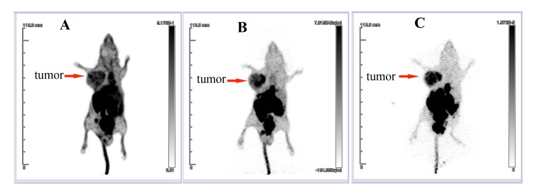
Fig 6. In vivo detection of glioma tumors using 18F-tLyP-1.
In vivo microPET/CT MIP images in the U87MG tumor model at 30 min (A), 60 min (B) and 120 min (C) after injection of 18F-tLyP-1. The uptake of 18F-tLyP-1 in the tumor is intense, while minimal uptake is seen in the brain. MIP: maximum-intensity image.

