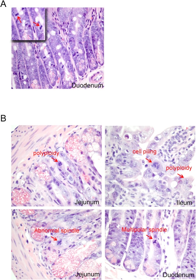
Fig 5. Histopathology of the small intestine of PF-7006-exposed mice.
(A) Vehicle-treatment showing diploid mitotic Figs (arrows) (inset) magnification of the image (n = 3). (B) Representative images of histological segments of the small intestine (duodenum, jejunum, ileum) from PF-7006 treated mice (n = 3) displaying abnormal mitotic Figs (arrows).

