
Figure 2. miR-193b inhibits motility and growth of ovarian cancer cells.
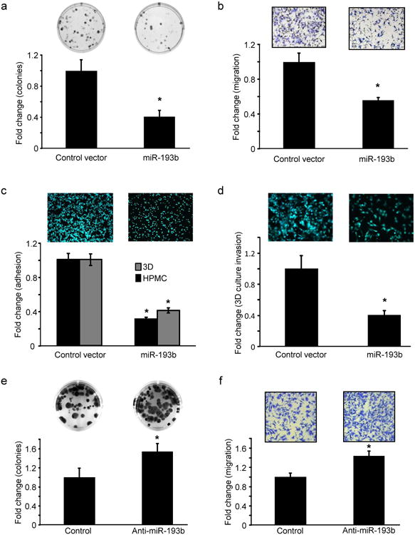
(a-d) miR-193b stable overexpression. (a) Colony formation. HeyA8 cells stably expressing miR-193b or vector controls were seeded in 6 well plates for a colony formation assay. Colonies were fixed after 2 weeks, stained and counted (mean ± SD; 3 independent experiments). (b) Migration. HeyA8 cells stably expressing miR-193b or control vector were seeded on 8 μm pore size inserts and allowed to migrate towards medium containing 10% FB. Migrated cells were fixed, imaged and quantified (mean±SD; 3 independent experiments). (c) Adhesion. HeyA8 GFP cells stably expressing miR-193b or control vector were seeded on the 3D culture model and allowed to adhere for 1 hour and quantified using a fluorescent plate reader (mean±SD; 3 independent experiments). (d) Invasion. HeyA8 GFP cells stably expressing miR-193b were allowed to invade through the 3D culture assembled in the upper chamber of 8 μm pore transwell inserts to assess their ability to invade through the surface of the omentum. The invaded HeyA8GFP cells were imaged and quantified (mean±SD; 3 independent experiments). (e-f) miR-193b inhibition. (e) Colony formation. HeyA8 cells were transfected with anti-miR-193b or negative control oligomers and a colony formation assay performed as described in (a) (mean±SD; 3 independent experiments). (f) Migration. HeyA8 cells were transfected with anti-miR-193b and used in a transwell migration assay (mean±SD; 3 independent experiments). * p<0.01, Students t test.