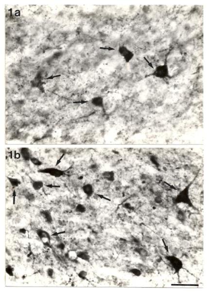
Fig. 1.
Photomicrographs of the ventrolateral portion of the central nucleus of the inferior colliculus showing the typical distribution of GAD-positive somata (arrows) in the Sprague-Dawley rat (a) and the GEPR (b). Note the increase in the numbers of small GAD-positive neurons in the GEPR. Scale bar = 25μm. Published with permission from Roberts et al. [10].

