
Fig. 5.
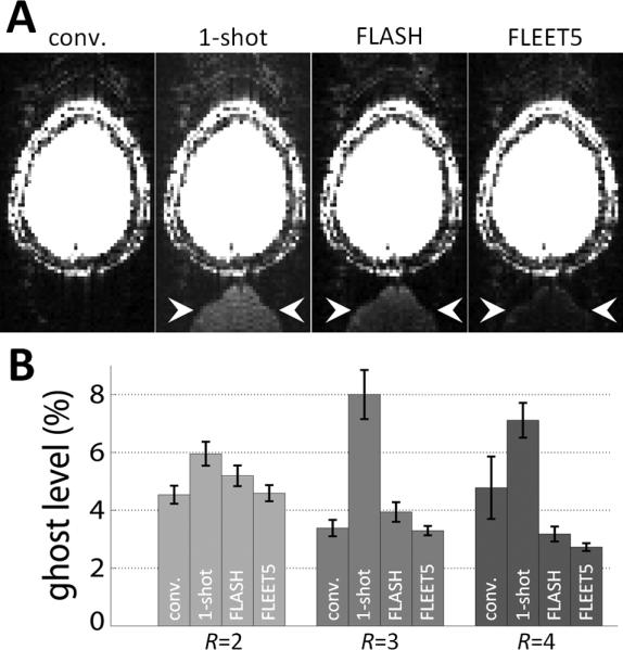
Dependence of residual aliasing on B0 effects. (A) Example images from R=3 accelerated images reconstructed using consecutive-slice ACS, single-shot EPI ACS (“1-shot”), FLASH-ACS, and FLEET5-ACS data from a 3T subject in the presence of a small, intentional shim offset to provoke differential distortion between the ACS data and the accelerated data. The pattern of the ghost in these examples is quite typical: a low ghost level is seen in the images reconstructed from the conventional consecutive-slice ACS data, while strong ghosting is seen when using the single-shot ACS data, and the ghosts seen when using FLASH-ACS data are prominent and spatially flat, whereas the ghosts seen when using the FLEET-ACS data are still visible but consist of a high-spatial-frequency edge artifact (ghost locations by arrowhead). The intensity scale in each panel is identical and is set to highlight the low-level ghosting. (B) A summary of the residual aliasing levels seen in an ROI outside of the brain across four 3T subjects. The ghost levels are clearly extreme in the images reconstructed from single-shot ACS data, and the lowest ghost levels are consistently seen in the images reconstructed with FLEET-ACS data.