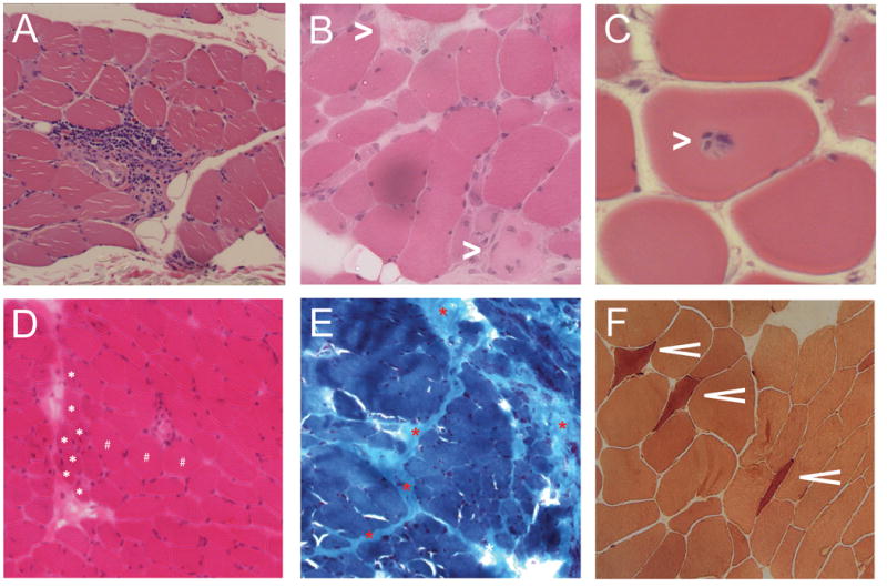
Figure 1.

Examples of histologic features from muscle biopsies of weak scleroderma patients. (A) Perivascular inflammation with extension into the endomysium (H&E stain). (B) Necrotic muscle fibers (white asterisks; H&E stain). (C) Focal invasion of an apparently healthy myofiber by lymphocytes (H&E stain). (D) Perifascicular atrophy (white asterisks indicate several atrophic fibers localized to the perifascicular area; H&E stain). (E) Prominent perimysial fibrosis (red asterisks; Gomori trichrome stain). (F) Numerous esterase-positive angular atrophic fibers suggesting an acute neurogenic process (esterase stain).
