
Figure 4.
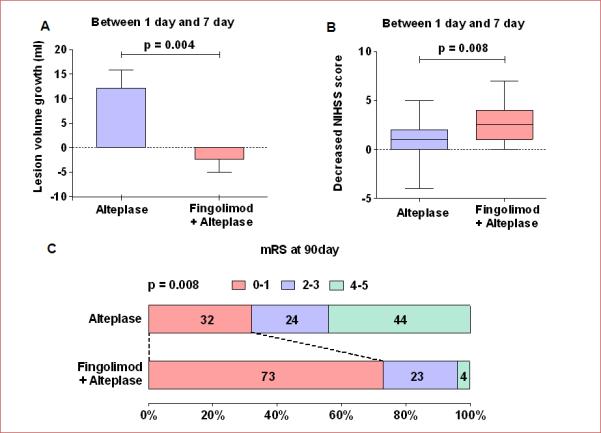
Lesion volume growth and clinical improvement from day 1 to day 7, as well as the probability of excellent recovery at 90 days. Panel A: Lesion volume growth from day 1 to day 7; lesion growth = Lesion volumes measured on Flair (day 7) minus Lesion volumes measured on Flair (day 1). Values are mean ± SE; comparisons were performed with independent t-tests. Panel B: Changes in the NIHSS score from day 1 to day 7; the horizontal line inside each box indicates the median, the top and bottom of the box indicate the interquartile range, the I bars indicate the 5th and 95th percentiles; comparisons were performed using the Mann-Whitney test; NIHSS = National Institutes of Health Stroke Scale. Panel C: Distribution of the degree of disability at day 90; comparisons were performed with Chi-squared test; mRS = modified Rankin Scale, Fin = fingolimod.