
Figure. 3.
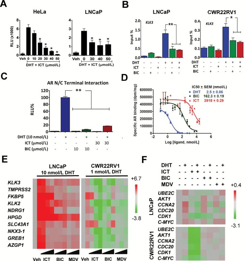
Effect of ICT on AR- or ARvs-signaling pathway. (A) AR transcriptional activity on consensus ARE was assessed in HeLa and LNCaP cells transfected with AR and ARE-Luc reporter genes. Luciferase activity was measured after exposure to either DMSO (Veh) or ICT with or without 10 nmol/l of DHT for 24h. (B) Chromatin immunoprecipitation of AR binding to the ARE of KLK3 gene. LNCaP and CWR22Rv1 cells were treated with 30 μmol/l of ICT or 10 μmol/l BIC alone or in combinations in the presence or absence of DHT for 2h. DNA fragments bound to AR were immunoprecipitated and analysed by the quantitative PCR. (C) AR NH2- and COOH-terminal (N–C) interactions were measured by a mammalian two-hybrid assay in CV-1 cells following exposure to indicated compounds for 24h. (D) Representative competition binding curves showing inhibition of 3H-DHT equilibrium binding to AR with increasing doses of unlabeled DHT, BIC and ICT in HeLa cells stably expressing AR. The IC50 values were determined using a one-site model using Graphpad Prism. (E) Full-length AR-regulated gene expression in LNCaP and CWR22Rv1 cells was assessed by quantitative RT–PCR following exposure to DMSO (Veh), 10, 30 and 50 µmol/l of ICT, BIC or MDV with or without DHT for 24h. (F) ARvs-regulated gene expression in LNCaP and CWR22Rv1 was assessed by quantitative RT–PCR following exposure to DMSO (Veh), 30 µmol/l of ICT, 10 µmol/l of BIC or MDV with or without 1 nmol/l DHT for 24h. Heatmaps show relative gene expression normalized to 18S rRNA. All data shown are mean ± SEM of at least three independent experiments. *P < 0.05; **P < 0.01.