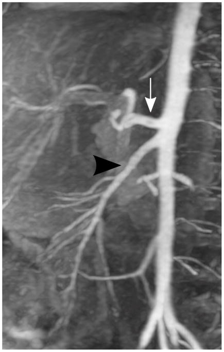
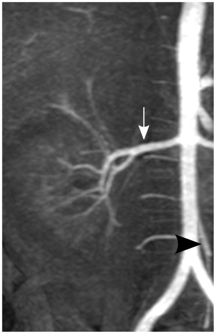
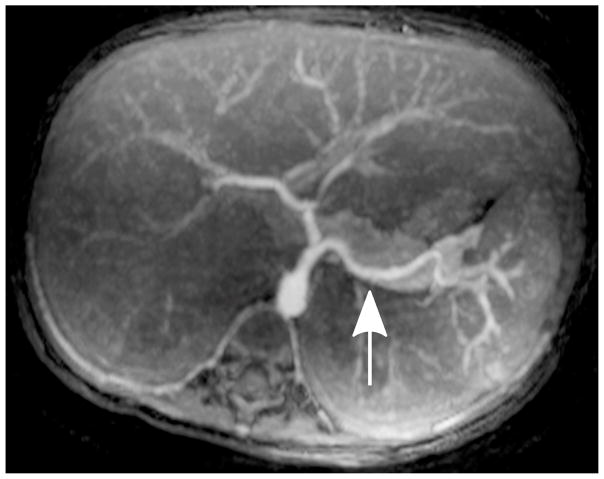
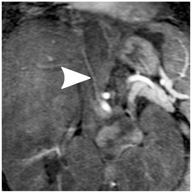
Fig. 2.
Representative MIP images of different abdominal arteries in the evaluation of a 3-year-old boy with endotracheal tube: (a) celiac artery (white arrow, scored 5 and 5 by readers 1 and 2, respectively) and superior mesenteric artery (black arrowhead, scored 5 and 5 by readers 1 and 2, respectively); (b) right renal artery (white arrow, scored 5 and 5 by readers 1 and 2, respectively) and inferior mesenteric artery (black arrowhead, scored 5 and 5 by readers 1 and 2, respectively); (c) splenic artery (white arrow, scored 5 and 4 by readers 1 and 2, respectively); (d) right phrenic artery (white arrowhead, scored 2 and 1 by readers 1 and 2, respectively)


